Sign-in, or Join to use this content. Join Restore

Atrial Rhythms ECG Interpretation

Overview

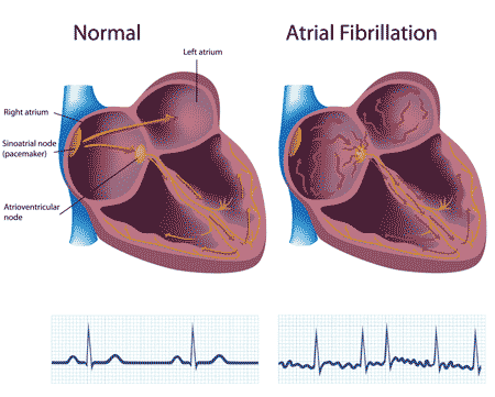
ventricular rhythm heart illustration

This page provides an introduction to atrial rhythms and links to training materials on this website.

Atrial rhythms originate in the atria, not from the SA node. The P wave's shape can be different from a normal sinus rhythm as the electrical impulse follows a different path. For a complete discussion of atrial rhythm ECG, use our atrial rhythms training module and our practice strips. Atrial rhythms categories:

  • Atrial Fibrillation (afib)
  • Atrial Flutter
  • Premature Atrial Complex
  • Multifocal Atrial Tachycardia
  • Supraventricular Tachycardia
  • Wandering Atrial Pacemaker
  • Wolff-Parkinson-White Syndrome
Sign-in, or Join to use this content. Join Restore

Atrial Rhythm Categories

Atrial Fibrillation

Atrial Fibrillation ECG tracing

Sites in the atria are firing very rapidly, between 400-600 bpm. These rapid pacemaking signals cause the atria to quiver. The ventricles beat at a slower rate because the AV node blocks some of the atrial impulses.

Atrial Flutter

Atrial Flutter ECG tracing

There are two types of atrial flutter. Type I (also called classical or typical) has a rate of 250-350 bpm. Type II (also called non-typical) are faster, ranging from 350-450 bpm. ECG tracings will show tightly spaced waves or saw-tooth shaped waveforms (F-waves).

Multifocal Atrial Tachycardia

Multifocal Atrial Tachycardia ECG tracing

During multifocal atrial tachycardia, several (non-SA) sites are creating impulses. The P waves will vary in shape and at least three different shapes can be observed. The PR Interval varies. Ventricular rhythm is irregular.

Premature Atrial Complex

Premature Atrial Complex ECG tracing

Premature atrial complex occurs when an ectopic site within the atria fires an impulse before the next impulse from the SA node. If the ectopic site is near the SA node, the P wave will often have a shape similar to a sinus rhythm. But this P wave will occur earlier than expected.

Supraventricular Tachycardia

Supraventricular Tachycardia ECG tracing

This term covers three types of tachycardia that originate in the atria, AV junction or SA node.

Wandering Atrial Pacemaker

 rhythm ECG tracing

Wandering atrial pacemaker is an irregular rhythm. In is similar to multifocal atrial tachycardia but the heart rate is under 100 bpm. P waves are present but will vary in shape.

Wolff-Parkinson-White Syndrome

 rhythm ECG tracing

Wolff-Parkinson-White Syndrome occurs when the impulse travels between the atria and ventricles via an abnormal path, called the bundle of Kent. The impulse, not being delayed by the AV node, can cause the ventricles to contract prematurely. ECG characteristics include a shorter PR Interval, longer QRS complex and a delta wave.
Click To Begin Atrial Rhythms Training Module


Lessons


Lessons
Click To Begin Atrial Rhythms Training Module


Lessons


Lesson #1: Rhythm Analysis Method - 312


Intro

The five steps of rhythm analysis will be followed when analyzing any rhythm strip.

  • Analyze each step in the following order.
  • Rhythm Regularity
  • Heart Rate
  • P wave morphology
  • P R interval or PRi
  • QRS complex duration and morphology

Step 1

Rhythm Regularity

ekg rhythm regularity
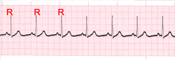
  • Carefully measure from the tip of one R wave to the next, from the beginning to the end of the tracing.
  • A rhythm is considered “regular or constant” when the distance apart is either the same or varies by 1 ½ small boxes or less from one R wave to the next R wave.

Step 2

Heart Rate Regular (Constant) Rhythms

regular ekg rhythm
  • The heart rate determination technique used will be the 1500 technique.
  • Starting at the beginning of the tracing through the end, measure from one R wave to the next R wave (ventricular assessment), then P wave to P wave (atrial assessment), then count the number of small boxes between each and divide that number into 1500. This technique will give you the most accurate heart rate when analyzing regular heart rhythms. You may include ½ of a small box i.e. 1500/37.5 = 40 bpm (don’t forget to round up or down if a portion of a beat is included in the answer).

Step 2 (Cont)

Heart Rate - Irregular Rhythms

irregular ekg rhythm
  • If the rhythm varies by two small boxes or more, the rhythm is considered “irregular”.
  • The heart rate determination technique used for irregular rhythms will be the “six-second technique”.
  • Simply count the number of cardiac complexes in six seconds and multiply by ten.

Step 3

P wave Morphology (shape)

ekg p wave tracing
  • Lead II is most commonly referenced in cardiac monitoring
  • In this training module, lead two will specifically be referenced unless otherwise specified.
  • The P wave in lead II in a normal heart is typically rounded and upright in appearance.
  • Changes in shape must be reported. This can be an indicator that the locus of stimulation is changing or the pathway taken is changing.
  • P waves may come in a variety of morphologies i.e. rounded and upright, peaked, flattened, notched, biphasic(pictured), inverted and even buried or absent!
  • Remember to describe the shape. This can be very important to the physician when diagnosing the patient.

Step 4

PR interval (PRi)

    PR interval regular
    Constant PR Interval

    PR interval irregular
    Variable PR Interval
  • Measurement of the PR interval reflects the amount of time from the beginning of atrial depolarization to the beginning of ventricular depolarization.
  • Plainly stated, this measurement is from the beginning of the P wave to the beginning of the QRS complex.
  • The normal range for PR interval is: 0.12 – 0.20 seconds (3 to 5 small boxes)
  • It is important that you measure each PR interval on the rhythm strip.
  • Some tracings do not have the same PRi measurement from one cardiac complex to the next. Sometimes there is a prolonging pattern, sometimes not.
  • If the PR intervals are variable, report them as variable, but note if a pattern is present or not.

Step 5

QRS complex

QRS Complex
  • QRS represents ventricular depolarization.
  • It is very important to analyze each QRS complex on the tracing and report the duration measurement and describe the shape (including any changes in shape).
  • As discussed in step 3, when referring to P waves, remember changes in the shape of the waveform can indicate the locus of stimulation has changed or a different conduction pathway was followed. It is no different when analyzing the QRS complex. The difference is that in step 3, we were looking at atrial activity. Now we are looking at ventricular activity.
  • Measure from the beginning to the end of ventricular depolarization.
  • The normal duration of the QRS complex is: 0.06 – 0.10 second


Lesson #2: Interpretation - 312


Introduction

  • The previous slides presented the five-steps of rhythm analysis. These five steps must be followed regardless of how simple of complex the tracing is you are reviewing.
  • The information gathered in these steps are telling a story.
  • The title of that story is the interpretation.

Atrial Dysrhythmias Types

The dysrhythmias in this category occur as a result of problems in the atria. These atrial dysrhythmias primarily affect the P wave. We will be discussing the following complexes and rhythms:

  • Premature Atrial Complexes (PAC’s)
  • Wandering Atrial Pacemaker
  • Multifocal Atrial Tachycardia
  • Atrial Flutter
  • Atrial Fibrillation


Lesson #3: Premature Atrial Complex


Intro to PAC

  • PAC's can occur for a number of different reasons i.e., diet, fatigue, stress, disease, ischemia to name a few.
  • Premature complexes frequently occur in bradycardic rhythms, but may occur almost any time.
  • PAC's occur when an early electrical impulse occurs from a location in the atria other than the SA node.

Intro to PAC 2

  • This early impulse causes an early cardiac complex which disrupts the underlying rhythm.
  • The locus of stimulation being different, results in a change in the morphology of the P wave.
  • PAC's can occur occasionally or frequently.
  • PAC's can be observed with or without a pattern
  • The P wave with PAC's will always be upright

ECG Analysis

atrial ecg image

Notice the following: the R to R interval is irregular, the fifth complex is early and the P wave on the early complex is a different shape.

ECG Practice Strip

atrial ecg image 2

Analyze this tracing using the five steps of rhythm analysis.

Show Answer
  • Rhythm: Irregular
  • Rate: 50
  • P wave: Upright & uniform (except early complexes - biphasic)
  • PR interval: 0.16 second
  • QRS: 0.08
  • Interpretation: Sinus Bradycardia with PAC's


Lesson #4: Wandering Atrial Pacemaker


Description

  • Rhythms are often named according to the origin of the electrical activity in the heart or the structure where the problem is occurring.
  • Wandering Atrial Pacemaker is aptly named due to the electrical impulses causing the atrial activity are moving or wandering.
  • These changes in the locus of stimulation affect the morphology of the P waves.

Analysis

wandering atrial pacemaker tracing
  • In Wandering Atrial Pacemaker, you must observe at least three different shaped P waves. No other changes in the tracing may be observed. The rhythm may or may not be regular.
  • The PR interval is often affected, but does not have to be.
  • The bottom line, is you must observe at least three different shaped P waves.

Practice Strip

wandering atrial pacemaker tracing

Analyze this tracing using the five steps of rhythm analysis.

Show Answer
  • Rhythm: Irregular
  • Rate: 50
  • P wave: Changing Shapes (3 or more)
  • PR interval: Variable
  • QRS: 0.08
  • Interpretation: Wandering Atrial Pacemaker


Lesson #5: Multifocal Atrial Tachycardia


Multifocal Atrial Tachycardia

  • Multifocal Atrial Tachycardia is just a faster version of Wandering Atrial Pacemaker. The criteria is the same as Wandering Atrial Pacemaker with the only difference being the heart rate exceeds 100 bpm.
  • These changes in the locus of stimulation within the atria affect the morphology of the P waves.
  • Remember, you must observe at least three different shaped P waves.
  • Due to the presence of irregular R to R intervals coupled with the changing P wave morphology, some people have confused this rhythm with Atrial Fibrillation.


Lesson #6: Atrial Flutter


Description

  • Atrial Flutter occurs when there is an obstruction within the atrial electrical conduction system.
  • Due to this impediment a series of rapid depolarizations occur.
  • These depolarizations may occur two, three, four or more times per QRS complex.
  • The AV node functions like a “gatekeeper” blocking the extra impulses until the ventricular conduction system is able to accept the impulse.
  • The impulse that is accepted will cause the QRS complex to occur.

Analysis

atrial flutter tracing
  • Each flutter wave represents atrial depolarization. This will be noted next to the P wave step in rhythm analysis. Instead of P waves, this tracing has “F” waves. No P waves mean there is no PR interval measurement.
  • When the tracing is interpreted, the ratio of F waves to each QRS complex will be documented along with the rhythm i.e. Atrial Flutter 4:1 (indicates 4 “F” waves to each QRS complex). Not all Atrial Flutter will have a regular rhythm. In that case just document and report your observations.

Practice Strip

atrial flutter ecg
  • Analyze this tracing using the five steps of rhythm analysis.
  • Compare your answers with the answers on the next slide.

Practice Strip Answers

atrial flutter ekg
  • Rhythm: Regular
  • Rate: Ventricles - 80, Atria - 320
  • P wave: "F" waves
  • PR interval: absent
  • QRS: 0.08
  • Interpretation: Atrial Flutter 4:1


Lesson #7: Atrial Fibrillation


Description

  • Atrial Fibrillation occurs when multiple electrical impulses occur within the atria. This chaotic electrical activity results in a chaotic wave form between the QRS complexes. P waves are absent. They are replaced by lower case "f" waves. No P waves means there is no PR interval measurement.
  • This rapid electrical activity overwhelms the AV node causing impulses to enter the ventricular conduction system at irregular points. This results in irregular R to R intervals.

Analysis

  • Not all fibrillatory waves are created equal. The "f" waves can be coarse (majority measure 3 mm or more) or can be fine (majority of waveforms measure less than 3 mm) to almost absent. Regardless always report your observations. Many times when a patient has "new onset" Atrial Fibrillation the patient will report with a heart rate of 160 bpm or more.

Mechanisms

atrial fibrillation tracing
  • When a patient experiences A-fib, the atria are not contracting as they normally would. They are just quivering. This absence of contraction of the atria can result in a loss of cardiac output anywhere from 15 - 30% due to the absence of "atrial kick". This is why the heart rate is so high. The body is trying to maintain homeostasis.
  • It will be impossible to determine the atrial rate. You will only be able to analyze and report the ventricular rate.
  • Atrial Fibrillation with a ventricular response in excess of 100 bpm is commonly referred to as Atrial Fibrillation with “rapid ventricular response” or "uncontrolled A-fib".

Practice Strip

atrial fibrillation ecg

Analyze this tracing using the five steps of rhythm analysis.

Show Answer
  • Rhythm: Irregular
  • Rate: Ventricles - 90, Atria - Unable to determine (UTD)
  • P wave: "f" waves
  • PR interval: absent
  • QRS: 0.08
  • Interpretation: Atrial Fibrillation


Lesson #8: Quiz: Test Questions - 312


Authors and Reviewers


Sources


?

An error has occurred. This application may no longer respond until reloaded. Reload 🗙