Parasternal Long Axis
Parasternal Long Axis
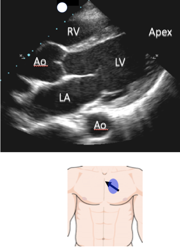
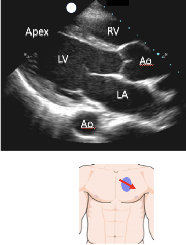
- Often most easily obtained
- Start high and move down lateral to sternum
Narration
Parasternal Long Axis
- Right ventricle (RV) anterior
-
Aorta (Ao) – root in center
- Descending aorta posterior
- Left atrium (LA) posterior
- Left ventricle (LV) and apex
-
Mitral valve well visualized
- Anterior/posterior leaflets
Narration
Parasternal Long Axis
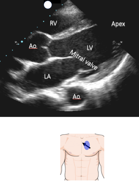
- Right ventricle (RV) anterior
-
Aorta (Ao) – root in center
- Descending aorta posterior
- Left atrium (LA) posterior
- Left ventricle (LV) and apex
-
Mitral valve well visualized
- Anterior/posterior leaflets

Narration
Parasternal Long Axis
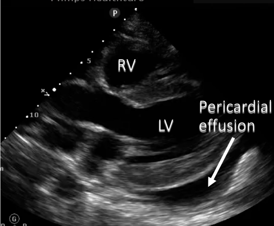
- Look posteriorly for effusion

Narration
Parasternal Long Axis
- Look posteriorly for effusion

Narration
Parasternal Long Axis
- Watch anterior leaflet of mitral valve to help estimate ejection fraction
Normal LV EF
Depressed LV EF

Narration
Parasternal Long Axis
- Watch anterior leaflet of mitral valve to help estimate ejection fraction
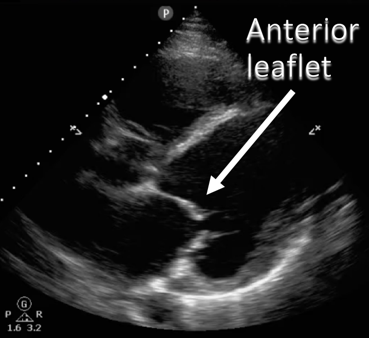
Normal LV EF
Depressed LV EF
